Radiology
-

-
 AED40.40
 -

Weir & Abrahams' Imaging Atlas of Human Anatomy 5E
Pages: 280,
Specialty: Radiology,
Publisher: Elsevier,
Publication Year: 2016,
Cover: Paperback
The perfect up-to-date imaging guide for a complete and 3-dimensional understanding of applied human anatomy Imaging is ever more integral to anatomy education and throughout modern medicine. Building on the success of previous editions, this fully revised fifth edition provides a superb foundation for understanding applied human anatomy, offering a complete view of the structures and relationships within the body using the very latest imaging techniques. It is ideally suited to the needs of medical students, as well as radiologists, radiographers and surgeons in training. It will also prove invaluable to the range of other students and professionals who require a clear, accurate, view of anatomy in current practice. * Fully revised legends and labels and over 80% new images - featuring the latest imaging techniques and modalities as seen in clinical practice * Covers the full variety of relevant modern imaging - including cross-sectional views in CT and MRI, angiography, ultrasound, fetal anatomy, plain film anatomy, nuclear medicine imaging and more - with better resolution to ensure the clearest anatomical views * Unique new summaries of the most common, clinically important anatomical variants for each body region - reflects the fact that around 20% of human bodies have at least one clinically significant variant * New orientation drawings - to help you understand the different views and the 3D anatomy of 2D images, as well as the conventions between cross-sectional modalities * Now a more compete learning package than ever before, with superb new BONUS electronic enhancements, including: - Labelled image 'stacks' - that allow you to review cross-sectional imaging as if using an imaging workstation - Labelled image 'slide-lines' - showing features in a full range of body radiographs to enhance understanding of anatomy in this essential modality - Self-test image 'slideshows' with multi-tier labelling - to aid learning and cater for beginner to more advanced experience levels - Labelled ultrasound videos - bring images to life, reflecting this increasingly clinically practiced technique - Questions and answers accompany each chapter - to test your understanding and aid exam preparation - 34 pathology tutorials - based around nine key concepts and illustrated with hundreds of additional pathology images, to further develop your memory of anatomical structures and lead you through the essential relationships between normal and abnormal anatomy
Weight: 0 KG
Learn MoreAED84.48 -
 AED110.19
 -
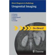
Urogenital Imaging, Dx-Direct Series
Pages: 258,
Specialty: Radiology,
Publisher: Thieme,
Publication Year: 2008,
Cover: Paperback,
Dimensions: 126x188x14mm
Dx-Direct gets to the point! Dx-Direct is a series of eleven Thieme books covering the main subspecialties in radiology. It includes all the cases you are most likely to see in your typical working day as a radiologist. For each condition or disease you will find the information you need -- with just the right level of detail. Dx-Direct gets to the point: * Definitions, Epidemiology, Etiology, and Imaging Signs * Typical Presentation, Treatment Options, Course and Prognosis * Differential Diagnosis, Tips and Pitfalls, and Key References All combined with high-quality diagnostic images. Whether you are a resident or a trainee, preparing for board examinations or just looking for a superbly organized reference: Dx-Direct is the high-yield choice for you! The series covers the full spectrum of radiology subspecialties including: Brain, Gastrointestinal, Cardiac, Breast, Genitourinal, Spinal, Head and Neck, Musculoskeletal, Pediatric, Thoracic, Vascular
Weight: 0.36 KG
Learn More -
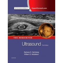
Ultrasound: The Requisites, 3rd Edition
Pages: 656,
Specialty: Radiology,
Publisher: Elsevier,
Publication Year: 2015,
Cover: Mixed media product,
Dimensions: 220.98x281.94x30.48mm
This best-selling volume in The RequisitesT Series provides a comprehensive introduction to timely ultrasound concepts, ensuring quick access to all the essential tools for the effective practice of ultrasonography. Comprehensive yet concise, Ultrasound covers everything from basic principles to advanced state-of-the-art techniques. This title perfectly fulfills the career-long learning, maintenance of competence, reference, and review needs of residents, fellows, and practicing physicians.
Weight: 2.06 KG
Learn MoreAED341.60 -
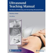
Ultrasound Teaching Manual, 3E
Pages: 126,
Specialty: Radiology,
Publisher: Thieme,
Publication Year: 2013,
Cover: Paperback,
Dimensions: 216x298x14mm
This workbook offers structured, course-like learning, and just like an instructor in an ultrasound course, it guides you systematically through the individual organ systems. The accompanying videos demonstrate basic anatomy for ultrasound, optimum transducer positioning, and the interaction between transducer position and monitor display, allowing you to experience the learning points in real time for a deeper, visual understanding. Highlights of the third edition: * Multiple-exposure photos demonstrate the dynamics of handling the transducer.* Triple-image sets clearly show transducer positioning, the ultrasound image, and an anatomic diagram of the site.* Numbered structures on the anatomic diagrams help you learn new information and test your retention at any time. The legend on the back-cover flap folds out for quick reference. Each structure is referred to by the same number throughout the book.* Numerous quiz images at the end of each chapter give you an opportunity to test your knowledge.* Physical principles are explained concisely with clear, accessible diagrams.* Various tips and tricks make it easier for beginners to get started. Ultrasound Teaching Manual is the perfect introduction to diagnostic ultrasound if you * are taking an ultrasound course and would like to prepare yourself systematically for this course or consolidate what you have learned* are a physician or student who wants to become familiar with diagnostic ultrasound in independent study; or* are a resident in internal medicine, radiology, surgery, gynecology, anesthesiology, or pediatrics who wants to solidify your ultrasound experience.
Weight: 0.58 KG
Learn MoreAED315.89 -
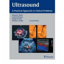
Ultrasound
Pages: 752,
Specialty: Radiology,
Publisher: Thieme,
Publication Year: 2007,
Cover: Hardback,
Dimensions: 218.44x284.48x38.1mm
Based on a popular course taught at the Radiological Society of North America's Annual Meeting, this book provides all the essential information for choosing the appropriate imaging examination and completing the imaging workup of a patient. Chapters are organized into parts according to the anatomical location of the clinical problems addressed. The authors guide the reader through the diagnostic evaluation, reviewing the indications for and the strengths and limitations of ultrasound imaging. Features: --Practical information on the usefulness of ultrasound, nonimaging tests, or other imaging modalities, such as CT and MR, for evaluating each clinical situation --Clear descriptions of symptoms and differential diagnosis --Nearly 1,300 images and photographs demonstrating key points --A new chapter on neonatal spinal cord anomalies Comprehensive and up-to-date, this edition is essential for ultrasonographers, radiologists, residents, physicians, nurses, and radiology assistants seeking the latest recommendations for the effective use of ultrasonography.
Weight: 2.54 KG
Learn MoreAED859.51 -

Ultrasonography in Urology
Pages: 192,
Specialties: Radiology, Urology,
Publisher: Thieme,
Publication Year: 2007,
Cover: Paperback,
Dimensions: 214x278x10mm
The second edition of Ultrasonography in Urology: A Practical Approach to Clinical Problems provides an up-to-date resource for the essential information needed for selecting the appropriate imaging examination and confidently completing the imaging workup of a patient. Recognized experts in the field provide the latest recommendations for clinical applications of ultrasound in urology. For each clinical problem, the authors guide the reader through the diagnostic evaluation, reviewing the indications for and the benefits and limitations of ultrasound imaging. Features: -Practical discussions of the usefulness of ultrasound, nonimaging tests, or other imaging modalities, such as CT and MR, for diagnosing such problems as flank pain, renal failure, acute scrotal pain, and more -Clear descriptions of symptoms and differential diagnosis -More than 400 high-quality images and photographs demonstrating key points This book will help ultrasonographers, radiologists, urologists, nephrologists, residents, physicians, nurses, and radiology assistants improve their techniques and optimize patient care.
Weight: 0.58 KG
Learn MoreAED352.62 -

Ultrasonography in Obstetrics and Gynecology
Pages: 272,
Specialty: Radiology,
Publisher: Thieme,
Publication Year: 2007,
Cover: Paperback,
Dimensions: 270x281x17mm
From diagnosing pelvic pain and bleeding, to the use of ultrasound in screening and treatments for ovarian cancer, infertility, and maternal complications of diabetes mellitus, this book covers the full spectrum of clinical applications for ultrasound in obstetrics and gynecology. The authors guide the reader through the diagnostic evaluation, reviewing the indications for and the strengths and limitations of ultrasound imaging, enabling clinicians to confidently choose the appropriate imaging examination for each clinical situation. Features: Practical information on the usefulness of ultrasound, non-imaging tests, or other imaging modalities, such as CT and MR Clear descriptions of symptoms and differential diagnosis Nearly 300 images and photographs demonstrating key points New chapter on amenorrhea in the adolescent or young adult This book is an essential resource for all ultrasonographers, radiologists, obstetricians, gynecologists, residents, physicians, nurses, and radiology assistants seeking to gain skill in the effective use of ultrasonography.
Weight: 0.82 KG
Learn More


