Radiology
-
Teaching Atlas of Urologic Imaging
Pages: 362,
Specialties: Radiology, Urology,
Publisher: Thieme,
Publication Year: 2009,
Cover: Hardback,
Dimensions: 220x284x20mm
Teaching Atlas of Urologic Imaging presents a case-based approach to selecting the multimodality imaging strategies for the most frequently encountered urologic disorders. The book provides comprehensive coverage of the latest imaging techniques with an emphasis on newer modalities such as CT intravenous pyelograms (CT-IVP) and MRI for the genitourinary system. Each case opens with a concise description of the clinical presentation, radiologic findings, diagnosis, and differential diagnosis. It then concludes with a detailed discussion of the background, clinical findings, pathology, imaging findings, treatment, and prognosis for that case, and pertinent references. Features: * Nearly 400 high-quality illustrations, including 47 in full color, demonstrate anatomy and pathology * Consistent format of each chapter enhances ease of use * Bulleted lists of differential diagnoses are ideal for rapid review Ideal for radiologists, urologists, and nephrologists, this book provides a quick reference for common imaging findings and the most appropriate imaging strategies for specific diseases. Its case-based format also makes it a valuable resource for residents preparing for board examinations.
Weight: 1.34 KG
Learn MoreBHD62.21 -
Teaching Atlas of Musculoskeletal Imaging
Pages: 800,
Specialty: Radiology,
Publisher: Thieme,
Publication Year: 2008,
Cover: Hardback,
Dimensions: 220x288x36mm
The latest addition to the popular Teaching Atlas series, Teaching Atlas of Musculoskeletal Imaging provides a complete overview of the most common manifestations of musculoskeletal disorders as well as the most important rare diseases. Internationally recognized authors guide the reader through multi-modality imaging approaches for 130 problems, which are grouped according to broad categories, including internal joint derangement, tumors, infection, avascular bone, trauma, arthritis, and prostheses. Each case provides concise descriptions of the presenting signs, radiologic findings, diagnosis, and differential diagnosis. Up-to-date information on musculoskeletal pathology and the current management strategies, including the latest interventional radiology techniques, make this atlas an outstanding reference for daily practice. Highlights: Essential information on the use of radiography, ultrasound, CT, and MRI enables clinicians to select the best combination of multiple imaging modalities for each case Bullet-point lists of "Pearls and Pitfalls" guide readers through diagnosis and help them avoid errors in image interpretation 900 images demonstrate key aspects of common and rare disease manifestations, providing an invaluable cross-reference tool for clinicians managing live cases Ideal for rapid reference and review, this atlas is an invaluable resource for clinicians and residents in radiology, orthopaedics, interventional musculoskeletal radiology, as well as those in musculoskeletal pathology, rheumatology, and sports medicine.
Weight: 2.42 KG
Learn MoreBHD67.48 -
Teaching Atlas of Abdominal Imaging
Pages: 544,
Specialty: Radiology,
Publisher: Thieme,
Publication Year: 2009,
Cover: Hardback,
Dimensions: 220.98x281.94x27.94mm
Teaching Atlas of Abdominal Imaging is a case-based reference covering the full spectrum of common and uncommon problems of the gastrointestinal and genitourinary tract encountered in everyday practice. The book organizes cases into sections based on the anatomic location of the problem. Each chapter provides succinct descriptions of clinical presentation, radiologic findings, diagnosis, and differential diagnosis for the case. The chapter then discusses the background for each diagnosis, clinical findings, common complications, etiology, imaging findings, treatment, and prognosis. Key features: * Succinct text and consistent presentation in each chapter enhance the ease of use * Practical discussion of all current imaging modalities * Nearly 550 high-quality images demonstrate key concepts * Bulleted lists of pearls and pitfalls at the end of each chapter highlight important points * An appendix with 64-slice protocols for various CT scans, such as dual-phase liver and pancreatic scans Ideal for both self-assessment and rapid review, this book is a valuable resource for radiologists, gastrointestinal and genitourinary radiologists, and fellows and residents in these specialties.
Weight: 1.7 KG
Learn MoreBHD52.03 -
Surgical Ultrasound
Pages: 445,
Specialties: Surgery, Radiology,
Publisher: Thieme,
Publication Year: 2007,
Cover: Hardback,
Dimensions: 204x274x30mm
This unique book is written specifically for surgeons. But that's not all. Edited by a surgeon and a gastroenterology specialist, it's relevant to both these disciplines. What's more, it's comprehensive and detailed: it covers everything from the technical basics, to emergency surgical ultrasound, to possible further diagnostic procedures. A truly practical guide Over 850 high-quality illustrations help to put the theory into practice. And explanatory drawings next to each ultrasound picture make it easy for even the inexperienced eye to interpret images. Clear, concise -- and easy to use Information is broken down into clear sections. Each section focuses on the surgical ultrasound of a specific organ system, and is written by a proven expert in the field. Topics include: -The acute abdomen -Thoracic and abdominal trauma -Endoscopic ultrasound -Doppler and color-coded Duplex ultrasound -Transplantation ultrasound -Intraoperative ultrasound
Weight: 1.58 KG
Learn MoreBHD36.19 -
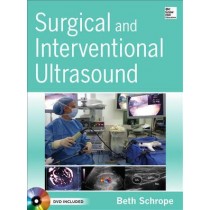
Surgical and Interventional Ultrasound
Pages: 274,
Specialties: Surgery, Radiology,
Publisher: McGraw-Hill,
Publication Year: 2013,
Cover: Mixed media product,
Dimensions: 224x278x18mm
All the guidance you need to enhance your understanding and clinical application of ultrasound Includes DVD with video of key techniques Surgical and Interventional Ultrasound offers a thorough survey of image-guided treatments in the OR, in the endoscopy suite, and at the bedside. This one-stop clinical companion spans virtually every kind of surgical and interventional specialty that utilizes ultrasound and delivers high-yield perspectives on using these techniques to ensure accurate clinical decision making. FEATURES: An all-in-one primer for ultrasound--packed with valuable how-to's and insights that take you through the basic exam and the full scope of interventions Essential content for residents that supplements training in surgery residency programs--from the Focused Assessment with Sonography for Trauma (FAST) exam, to intraoperative ultrasound and ultrasound-guided procedures such as breast biopsy or radiofrequency ablation Up-to-date, multidisciplinary focus on surgical and interventional ultrasound covers the array of procedures for which ultrasound is increasingly utilized Full-color illustrations with hundreds of ultrasound images Valuable opening chapter on the physics of ultrasound, which enables better quality images and a better understanding of image interpretation Important chapter on advanced technologies highlights 3D ultrasound imaging and contrast ultrasound, drawing attention to their safe and effective implementation in surgical practice Emphasis on ultrasound-guided anesthesia explains how ultrasound can enhance the precision of regional anesthetic procedures Instructive companion DVD features clips of key diagnostic and interventional techniques
Weight: 1.02 KG
Learn MoreBHD67.48 -

Succeeding in the FRCR Part 1 Exam (Physics Module), 2e
Pages: 264,
Specialties: Radiology, UK - FRCR,
Publisher: NA,
Publication Year: 2011,
Cover: Paperback
Weight: 0.46 KG
Learn MoreBHD6.79 -
Study Guide and Laboratory Exercises for Technology for Diagnostic Sonography
Pages: 204,
Specialty: Radiology,
Publisher: Elsevier,
Publication Year: 2012,
Cover: Paperback,
Dimensions: 215.9x271.78x5.08mm
Gain a firm foundation for sonography practice! Corresponding to the chapters in Hedrick's "Technology for Diagnostic Sonography", this study guide focuses on basic concepts to help you master sonography physics and instrumentation. It includes laboratory exercises designed to teach you how to operate a scanner, and comprehensive review questions allow you to assess your knowledge. Not only will you learn the theoretical knowledge that is the basis for ultrasound scanning, but also the practical skills necessary for clinical practice.
Weight: 0.45 KG
Learn MoreBHD23.00 -

Stoller's Atlas of Orthopaedics and Sports Medicine
Pages: 1040,
Specialty: Radiology,
Publisher: Wolters Kluwer,
Publication Year: 2008,
Cover: Hardback,
Dimensions: 241.3x307.34x48.26mm
Using 1,298 full-color anatomic drawings and 230 3-Tesla MR normal anatomy images, this atlas provides a detailed view of the intricacies of musculoskeletal anatomy. Dr. Stoller, through extensive cadaver dissections and imaging, has developed and proven new concepts on many musculoskeletal injuries, including hip impingement and patterns of meniscal tears. In this atlas, he provides radiologists, orthopaedists, and other specialists with the anatomic knowledge needed to accurately diagnose musculoskeletal injuries. Muscles are shown in great detail, including origins and insertions. Skeletal structures are shown in relation to muscles, tendons, nerves, and ligaments. Clear legends describe the function and directional movement of muscles. Illustrations show both normal anatomy and mechanisms of injury, and Pearls and Pitfalls sections reinforce critical information.
Weight: 3.95 KG
Learn More -

Step by Step MRI with CD-ROM
Pages: 300,
Specialty: Radiology,
Publisher: Jaypee,
Publication Year: 2008,
Cover: Mixed media product,
Dimensions: 121.92x172.72x12.7mm
Weight: 0.32 KG
Learn More -
 BHD74.27


