Radiology
-

300 Single Best Answers for the Final FRCR Part A
Pages: 160,
Specialties: Radiology, UK - FRCR,
Publisher: JP Medical,
Publication Year: 2010,
Cover: Paperback,
Dimensions: 226.06x289.56x63.5mm
300 Single Best Answers for the Final FRCR Part A provides 300 practice MCQs, in the new style single best answer format, for candidates preparing for Final FRCR examinations. The book is organised into six chapters that reflect the format of the exam. Each chapter comprises 50 MCQs and an answer section that provides a detailed rationale for the correct response. Key Points * Q&As mirror the new Single Best Answer format adopted by the RCR in 2009: few other books available in this new format * Detailed rationales provided for every single question, so candidates understand why the correct answer is right
Weight: 4.26 KG
Learn MoreKWD12.00 -
A-Z of Abdominal Radiology
Pages: 366,
Specialty: Radiology,
Publisher: Cambridge,
Publication Year: 2009,
Cover: Paperback,
Dimensions: 154x234x24mm
A-Z of Abdominal Radiology provides a concise, easily accessible radiological guide to the imaging of the common disorders of the abdomen and pelvis. Organised by A-Z, each entry gives easy access to the key clinical features of the condition. Section 1 reviews the relevant radiological anatomy of the abdomen and pelvis. This is followed by over 80 abdominal disorders, listing characteristics, clinical features, radiological features and relevant clinical management. Each disorder is highly illustrated to aid diagnosis. A-Z of Abdominal Radiology is an invaluable quick reference for the busy clinician and aide memoir for exam revision in both medicine and radiology.
Weight: 0.78 KG
Learn MoreKWD19.49 -
Abdominal Imaging, 2-Volume Set
Pages: 1600,
Specialty: Radiology,
Publisher: Elsevier,
Publication Year: 2010,
Cover: Mixed media product,
Dimensions: 251.46x304.8x129.54mm
"Abdominal Imaging", a title in the "Expert Radiology Series", edited by Drs. Dushyant Sahani and Anthony Samir, is a comprehensive 2-volume reference that encompasses both GI and GU radiology. It provides richly illustrated, advanced guidance to help you overcome the full range of diagnostic, therapeutic, and interventional challenges in abdominal imaging and combines an image-rich, easy-to-use format with the greater depth that experienced practitioners need. Online access at expertconsult.com allows you to rapidly search for images and quickly locate the answers to any questions.
Weight: 5.35 KG
Learn MoreKWD91.17 -

Abdominal Ultrasound: Step by Step 3E
Pages: 352,
Specialty: Radiology,
Publisher: Thieme,
Publication Year: 2016,
Cover: Paperback,
Dimensions: 196x269x18mm
The third edition of this practical reference guide has been updated with a modern, visually attractive design and expanded content. The book is ideal for healthcare professionals with little or no experience in administering and interpreting abdominal ultrasound examinations. It is practice-oriented and structured in a way that allows readers with varying degrees of ultra-sonography knowledge to utilize the material according to their individual experience and needs. Each chapter includes a systematic, detailed description of the anatomy involved in the ultrasound examination, with easy-to-digest steps that follow standardized routine and protocol. That straight-forward approach, coupled with more than 1,000 high-quality images and illustrations, enables hands-on learning, yielding the ability to assimilate these techniques quickly and adeptly. This is a stellar resource that provides the requisite tools to locate and display the anatomical structure being tested, position and move the transducers accurately, describe and interpret the findings correctly, and differentiate key findings from the many image artifacts that typically occur. Key Highlights: * In-depth discussion of organ boundaries, organ details, anatomical relationships, potentially abnormal findings, tips, and clearly defined learning objectives * Anatomical drawings incorporate a "sliced 3-D" view that show how the structures are displayed by the sector-shaper beam * Each chapter includes a series of images replicating the 3-D impression that results from the transducer moving across the body * Schematic drawings illustrate the ultrasound images, including a body marker that shows the transducer position * The "sono-consultant": a systematic guide to evaluating ultrasound findings and establishing a differential diagnosis This step-by-step guide is an invaluable, pragmatic resource to have on hand while performing abdominal ultrasound on the patient. In-depth but concise, this is an essential teaching guide for medical students, residents, technicians, and physicians who need to learn and master these examination techniques.
Weight: 0.96 KG
Learn MoreKWD24.89 -
Abdominal X-rays for Medical Students
Pages: 128,
Specialty: Radiology,
Publisher: Wiley,
Publication Year: 2015,
Cover: Paperback,
Dimensions: 212x274x18mm
Abdominal X-rays for Medical Students is a comprehensive resource offering guidance on reading, presenting and interpreting abdominal radiographs. Suitable for medical students, junior doctors, nurses and trainee radiographers, this brand new title is clearly illustrated using a unique colour overlay system to present the main pathologies and to highlight the abnormalities in abdomen x-rays. Abdominal X-rays for Medical Students: * Covers the key knowledge and skills necessary for practical use * Provides an effective and memorable way to analyse and present abdominal radiographs - the unique 'ABCDE' system as developed by the authors * Presents each radiograph twice, side by side: the first as seen in the clinical setting, and the second with the pathology clearly highlighted * Includes self-assessment to test knowledge and presentation technique With a systematic approach covering both the analysis of radiographs and next steps mirroring the clinical setting and context, Abdominal X-rays for Medical Students is a succinct and up-to-date overview of the principles and practice of this important topic.
Weight: 0.38 KG
Learn MoreKWD10.80 -

Abdominal X-Rays Made Easy, International Edition, 2nd Edition
Pages: 228,
Specialties: Radiology, Surgery,
Publisher: Elsevier,
Publication Year: 2006,
Cover: Paperback,
Dimensions: 124x184x14mm
This lively and entertaining manual on how to interpret abdominal radiographs will be invaluable to all medical students and junior doctors and has been written by a practising radiologist with many years' experience of teaching the subject. It outlines the few simple rules you need to follow, then explains how to sort out the initial and apparently overwhelming jumble of information which constitutes the abdominal X-ray. Knowledge of its contents will provide a secure base for tackling exams and the subsequent challenges of clinical practice.
Weight: 0.28 KG
Learn MoreKWD4.50 -
Accident and Emergency Radiology: A Survival Guide, 3e
Pages: 384,
Specialty: Radiology,
Publisher: Elsevier,
Publication Year: 2014,
Cover: Paperback,
Dimensions: 142x222x20mm
This pocket book is written primarily for doctors with little or no experience in the accident and emergency department and who are faced with the problem of radiological interpretation when no other help is readily at hand. Step-by-step methods for the assessment of radiographs help to answer the question `these look normal to me, but how can I be sure that I am not overlooking a subtle but important abnormality`. The book is liberally illustrated with high quality, well-annotated radiographs which assist the reader in the interpretation of abnormal/normal radiographic appearances. The focus is on common areas of injury with the emphasis being placed on the detection of those abnormalities that are most commonly overlooked or misinterpreted. Each chapter deals with the basic radiographs required, important anatomy, normal variants, a system for inspecting suggested views, types of injury and ends with a summary of key points.
Weight: 0.68 KG
Learn More -

AIDS to Part 1 FRCR
Pages: 144,
Specialties: Radiology, UK - FRCR,
Publisher: NA,
Publication Year: 1988,
Cover: Paperback,
Dimensions: 140x220mm
Weight: 0.18 KG
Learn MoreKWD14.10 -

An Introduction to Radiography
Pages: 376,
Specialty: Radiology,
Publisher: Elsevier,
Publication Year: 2009,
Cover: Paperback,
Dimensions: 188x246x18mm
This book provides a solid foundation in radiography for first year degree students by giving an overview of the basic principles and inspiring them to explore further the concepts presented. It also covers the core knowledge and standards for professional practice in sufficient depth to enable assistant practitioners to pass their NVQ examinations, practise their skills effectively and provide good patient care. show more
Weight: 0.82 KG
Learn More -
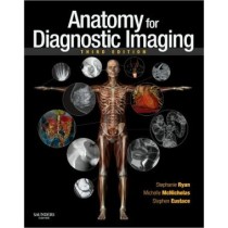
Anatomy for Diagnostic Imaging, 3e
Pages: 384,
Specialty: Radiology,
Publisher: Elsevier,
Publication Year: 2010,
Cover: Paperback,
Dimensions: 218x274x18mm
This book covers the normal anatomy of the human body as seen in the entire gamut of medical imaging. It does so by an initial traditional anatomical description of each organ or system followed by the radiological anatomy of that part of the body using all the relevant imaging modalities. The third edition addresses the anatomy of new imaging techniques including three-dimensional CT, cardiac CT, and CT and MR angiography as well as the anatomy of therapeutic interventional radiological techniques guided by fluoroscopy, ultrasound, CT and MR. The text has been completely revised and over 140 new images, including some in colour, have been added. A series of 'imaging pearls' have been included with most sections to emphasise clinically and radiologically important points. The book is primarily aimed at those training in radiology and preparing for the FRCR examinations, but will be of use to all radiologists and radiographers both in training and in practice, and to medical students, physicians and surgeons and all who use imaging as a vital part of patient care. The third edition brings the basics of radiological anatomy to a new generation of radiologists in an ever-changing world of imaging.
Weight: 0.98 KG
Learn MoreKWD22.49


